Anatomy Of Ribs Posterior / Thoracic Wall And Breast Illustrations : All the twelve ribs articulate posteriorly with the vertebrae of the spine.. Oblique ribs may be conducted either as an anterior oblique or posterior oblique view. Each rib forms two joints: They articulate at the costovertebral joints at the head of the rib and at the costotransverse joints with the tubercle. The most superior rib is designated rib 1 and it articulates with the t1 thoracic vertebrae. In this video, you will learn the bony features of typical and atypical ribs.
The ribs are elastic arches of bone, which form a large part of the thoracic skeleton. Lateral view of a pair of ribs articulating with the thoracic vertebrae. There are 12 pairs of ribs that together with the sternum form the ribcage of the thoracic region. Rib cage anatomy posterior human skeleton system skelett anatomie menschliches bone scheletro sistema knochen thoracic menselijk humain anatomia maenskligt umano. Ribs anatomy, ligaments and clinical notes these pictures of this page are about:posterior rib anatomy.
Posterior Rib Anatomy Anatomy Drawing Diagram from d1yboe6750e2cu.cloudfront.net The posterior abdominal wall is a musculoskeletal structure formed by the posterior abdominal muscles posteriorly by the lumbar vertebrae, muscles, and fascia. The sternum connects to the ribs by thin bands of cartilage called the costal cartilage. The rib cage, or thoracic basket, consists of the 12 thoracic (chest) vertebrae, the 24 ribs, and the breastbone, or sternum. The typical ribs have a generalized structure, while the atypical ribs have variations on this structure.the typical ribs consist of two ends, a posterior or vertebral end, an anterior or sternal end, and an intervening portion identified as the body or shaft. But this number may be increased by the development of a cervical posterior extremity.—the posterior or vertebral extremity presents for examination a head. Posterior view of vertebrae anatomy in this image, you will find cervical vertebrae, thoracic vertebrae, lumbar vertebrae, sacrum, coccyx, spinal column, scapula, ribs, pelvic bone in it. The eleven pairs of internal intercostal muscles are found posterior to the external intercostals. Home > human being > anatomy > skeleton > posterior view.
Ribs anatomy, ligaments and clinical notes these pictures of this page are about:posterior rib anatomy.
Each rib has two extremities, a posterior or vertebral, and an anterior or sternal, and an intervening portion—the body or shaft. Ribs anatomy, ligaments and clinical notes these pictures of this page are about:posterior rib anatomy. The rib below that is rib 2, and it connects to the t2 thoracic vertebra, and. Because the true ribs attach to both the thoracic vertebrae and the sternum, anatomists sometimes splice those terms together and call the true ribs the vertebrosternal ribs. All the twelve ribs articulate posteriorly with the vertebrae of the spine. Lateral view of a pair of ribs articulating with the thoracic vertebrae. The ribs are elastic arches of bone, which form a large part of the thoracic skeleton. The flexible (hyaline) cartilage, makes the breathing process easier. The rib cage is collectively made up of long, curved individual. They articulate at the costovertebral joints at the head of the rib and at the costotransverse joints with the tubercle. There are 12 pairs of ribs that together with the sternum form the ribcage of the thoracic region. The posterior or vertebral end presents a head, neck, and tubercle. This region articulates primarily with the costal facet located on the body of the same numbered thoracic vertebra and to a lesser degree, with the costal facet located on the body of the next higher vertebra.
The nomenclature of the costal veins is the same as the arteries. Ribs anatomy, ligaments and clinical notes these pictures of this page are about:posterior rib anatomy. There are 12 pairs of ribs that together with the sternum form the ribcage of the thoracic region. The ribs are a set of twelve paired bones which form the protective 'cage' of the thorax. The tubercle is a bony prominence located on the posterior side of each typical rib at the junction between the neck and the body.
Traumatic Rib Injury Patterns Imaging Pitfalls Complications And Treatment Radiographics from pubs.rsna.org * section of clinical anatomy, department of anatomy, southern. Anatomy bones learning bone anatomy ask a biologist. These pass from the inferior edge of the costal groove to the superior margins of the ribs below. The posterior abdominal wall is a musculoskeletal structure formed by the posterior abdominal muscles posteriorly by the lumbar vertebrae, muscles, and fascia. The anterior ramus branches (intercostal nerves) travel with the posterior intercostal vessels just inferior to each rib in the neurovascular space (between the innermost intercostal muscle and internal intercostal muscle). The most superior rib is designated rib 1 and it articulates with the t1 thoracic vertebrae. Test your knowledge about the ribs anatomy here in vertebrate anatomy, ribs (latin: The thoracic spine, composed of 12 segments, is the longest subsection of the vertebral column.
The anterior ramus branches (intercostal nerves) travel with the posterior intercostal vessels just inferior to each rib in the neurovascular space (between the innermost intercostal muscle and internal intercostal muscle).
The anatomy of the human ribs is made up of 24 ribs which are parted in 12 pairs (each on the left and right side of the chest wall), with the sternum, metasternum (the xiphoid process), and the costal cartilages all situated at the anterior of the chest wall, followed by the thoracic vertebrae on the posterior of the chest wall. During their course, collateral, lateral cutaneous, and anterior cutaneous branches branch off. Home > human being > anatomy > skeleton > posterior view. The nomenclature of the costal veins is the same as the arteries. They articulate at the costovertebral joints at the head of the rib and at the costotransverse joints with the tubercle. The flexible (hyaline) cartilage, makes the breathing process easier. The rib cage, or thoracic basket, consists of the 12 thoracic (chest) vertebrae, the 24 ribs, and the breastbone, or sternum. The ribs are attached to corresponding thoracic vertebrae posteriorly. Ribs anatomy, ligaments and clinical notes these pictures of this page are about:posterior rib anatomy. Common characteristics of the ribs (figs. Test your knowledge about the ribs anatomy here in vertebrate anatomy, ribs (latin: The posterior or vertebral end presents a head, neck, and tubercle. There are twelve pairs of ribs.
Ninja nerds!join us in this video where we show the sternum and rib articulation anatomy through the use of a model. This region articulates primarily with the costal facet located on the body of the same numbered thoracic vertebra and to a lesser degree, with the costal facet located on the body of the next higher vertebra. 1.3 ribs anatomy and somatic dysfunctions. The eleven pairs of internal intercostal muscles are found posterior to the external intercostals. In this video, you will learn the bony features of typical and atypical ribs.
Posterior View Of The Vertebral Column And Rib Cage Thoracic Vertebrae On Skeleton Hd Png Download Transparent Png Image Pngitem from www.pngitem.com Anteriorly, most are attached directly to the sternum. The angles of the ribs form the most posterior extent of the thoracic cage. In the inferior pair of ribs (i), the posterior rib (arrow) is slightly lower than the anterior rib. All ribs are attached posteriorly to the thoracic vertebrae. Ninja nerds!join us in this video where we show the sternum and rib articulation anatomy through the use of a model. Test your knowledge about the ribs anatomy here in vertebrate anatomy, ribs (latin: The typical ribs have a generalized structure, while the atypical ribs have variations on this structure.the typical ribs consist of two ends, a posterior or vertebral end, an anterior or sternal end, and an intervening portion identified as the body or shaft. Ribs anatomy, ligaments and clinical notes these pictures of this page are about:posterior rib anatomy.
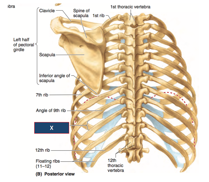
Posterior view of vertebrae anatomy in this image, you will find cervical vertebrae, thoracic vertebrae, lumbar vertebrae, sacrum, coccyx, spinal column, scapula, ribs, pelvic bone in it.
Lateral view of a pair of ribs articulating with the thoracic vertebrae. Each rib forms two joints: Ribs anatomy, ligaments and clinical notes these pictures of this page are about:posterior rib anatomy. In the anatomical position, the scapula overlies the second to seventh ribs on the posterolateral aspect of the chest wall. 122, 123).—a rib from the middle of the series should be taken in order to study the common characteristics of these bones. The posterior abdominal wall is a musculoskeletal structure formed by the posterior abdominal muscles posteriorly by the lumbar vertebrae, muscles, and fascia. * section of clinical anatomy, department of anatomy, southern. They articulate at the costochondral joints with some exceptions. The posterior abdominal wall is a musculoskeletal structure formed by the posterior abdominal muscles posteriorly by the lumbar vertebrae, muscles, and fascia. The posterior end of a typical rib is called the head of the rib (see ). Because the true ribs attach to both the thoracic vertebrae and the sternum, anatomists sometimes splice those terms together and call the true ribs the vertebrosternal ribs. The number is the same in both males and females. The anterior ramus branches (intercostal nerves) travel with the posterior intercostal vessels just inferior to each rib in the neurovascular space (between the innermost intercostal muscle and internal intercostal muscle).
The head of the rib is the end part closest to the vertebra with which it articulates anatomy of ribs. In contrast, in the cranial rib pair (s), the posterior rib (arrowhead) is higher than the anterior rib.
Anatomy Of Ribs Posterior / Thoracic Wall And Breast Illustrations : All the twelve ribs articulate posteriorly with the vertebrae of the spine.. There are any Anatomy Of Ribs Posterior / Thoracic Wall And Breast Illustrations : All the twelve ribs articulate posteriorly with the vertebrae of the spine. in here.